
You’ve scheduled your mammogram! Great work!
In this post, we’ve included information about this type of screening for breast cancer and a step-by-step look at what it’s like to get one. We want every woman to know what to expect during a mammogram!
Let's get started.
What Age Should You Get a Mammogram?
Women at average risk for developing breast cancer should begin getting annual mammograms at age 40, and earlier if breast cancer runs in their family. If there is a history of breast cancer in a first-degree relative (i.e. your mother, sister, or grandmother), then you should begin screening 10 years prior to their age at diagnosis or at age 40, whichever comes first.

Additionally, certain groups have a high prevalence of more aggressive cancers at younger ages. For instance, African American women have a high incidence of triple-negative breast cancer, which in addition to being aggressive, is harder to treat and has worse outcomes. Ashkenazi Jewish women have much higher rates of breast cancer because of genetic mutations. Speak to your doctor about whether you should start screening earlier than 40.
What to Expect During a Mammogram
The Day of Your Mammogram
When you arrive at the breast care center or location, you’ll check in at the front desk. Check-in includes verifying your name and personal information. If you have insurance, they’ll collect your insurance card and ask you to complete forms for registration. Once you are registered, a radiology technologist will escort you to get your mammogram.
Right Before Your Mammogram
Before the exam, you’ll be asked to change into a medical gown. The technologist will place radiodense stickers on your skin on top of any moles, scars, or raised skin lesions so the radiologist is aware of them while interpreting your images. This does not hurt at all. It is also important to provide the technologist with your medical history especially pertaining to your breasts. This will also help the radiologist in interpreting your images.
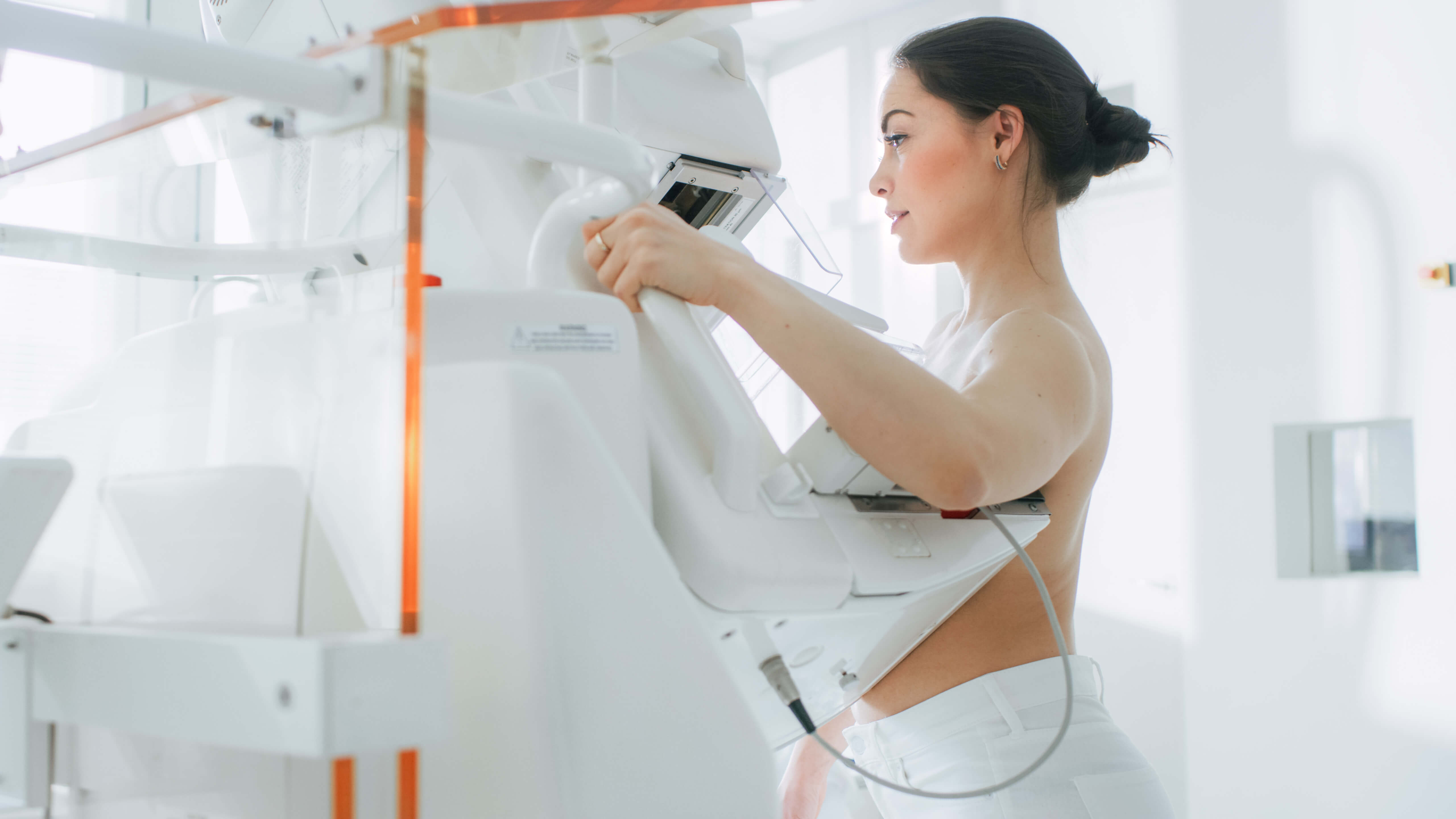
The Exam Itself
A mammogram machine is a tall standing structure that uses x-rays to image the breast tissue. The technologist will position your breasts in the mammogram machine and apply compression or pressure. The compression can feel somewhat uncomfortable at first but you are only required to be in compression for a few seconds at a time while the images are being taken. Compression is important because it decreases the radiation dose to the patient and decreases the time to obtain the images.
Once you are in an optimal position, the technologist will leave the room. She will then take multiple images from behind a glass wall. The radiation in a mammogram is very minimal and nearly inconsequential to each patient, however, because the technologist performs this process on many patients throughout the day, she needs to protect herself from the exposure.
Once the images are done, your exam is complete. You can change into your clothes and go on with the rest of your day.
After Your Mammogram
After the images are taken, your mammogram will be interpreted by a radiologist who will generate a report with the results. Interpretation of the mammogram includes a finding of any new or dominant masses, asymmetries, or calcifications. If a mammogram is completely clear and has no findings to report, that is considered a BI-RADS category 1 or negative. If there are findings to report that are stable or do not require any further evaluation, such as metallic biopsy markers from prior biopsies or typically benign calcifications, then it is reported as a category 2 or benign. If there is a finding that needs further evaluation or work-up, then it is reported as category 0 for further evaluation.
The radiologist interpreting your images will also evaluate breast density. Breast density is the amount of connective tissue (or fibrous tissue) versus fatty tissue in your breasts. There are four categories of breast density depending on how much fibrous tissue to fatty tissue is present within the breast. On the mammogram, the fibrous breast tissue appears gray or white, and fatty tissue appears black or dark gray. The four categories of breast density are:
1. Almost entirely Fatty
2. Scattered fibroglandular density
3. Heterogeneously dense
4. Extremely dense.
The sensitivity of mammograms decreases with increasing breast density. Therefore, supplemental screening with ultrasound or MRI should be considered in those with a category 3 or 4 breast density.

Your Mammogram Results and Report
After the test, you’ll receive a report in the mail, or it will be uploaded to a medical portal. The results of the report should be mailed within 30 days, often sooner. The doctor who ordered the mammogram (either your primary care doctor or gynecologist) will also receive a copy of the same report.
If your screening mammogram report is given a BIRADS category 0 then you will be asked to return for further evaluation with either a diagnostic mammogram that includes specialized views, an ultrasound, or both. Neither of these tests is invasive.
Additionally, the report will often include information about your breast density, based on your mammogram images. There are 38 states plus the District of Columbia that require the reporting of breast density to patients. In 11 states insurance companies must cover the cost of additional tests for women with dense breasts. Additionally, federal law will soon require all women in the United States to be notified about breast density following a mammogram.

Make sure to learn your breast density and if you have dense breasts, request an ultrasound in addition to your mammogram.
Screen Every Year
It is very important to keep up with yearly screening exams to maximize your chances of finding early curable cancers and to establish what is normal for each individual. The most helpful tool for a radiologist when evaluating your screening mammogram is a comparison to prior examinations. Baseline screening mammograms for women considered at average risk of getting breast cancer should begin at age 40 and should be performed yearly. If you have prior examinations from an outside institution, it is highly recommended to obtain those studies for comparison. This can, potentially, help avoid breast cancer.
Breast cancer is most treatable when caught early, which is why annual mammograms are the key to saving lives.
Photo by Dainis Graveris on Unsplash

About Dr. Mansi Vaishnav
Mansi was raised in New Orleans, Louisiana and attended Louisiana State University, where she obtained her undergraduate degree in basic sciences in 2009. She obtained her MD summa cum laude from the American University of Antigua. She went to train in diagnostic radiology at the University of Mississippi medical center and graduated in June 2020. In her free time she enjoys cooking, exercising, and spending time with her family.